What is an Echocardiogram?
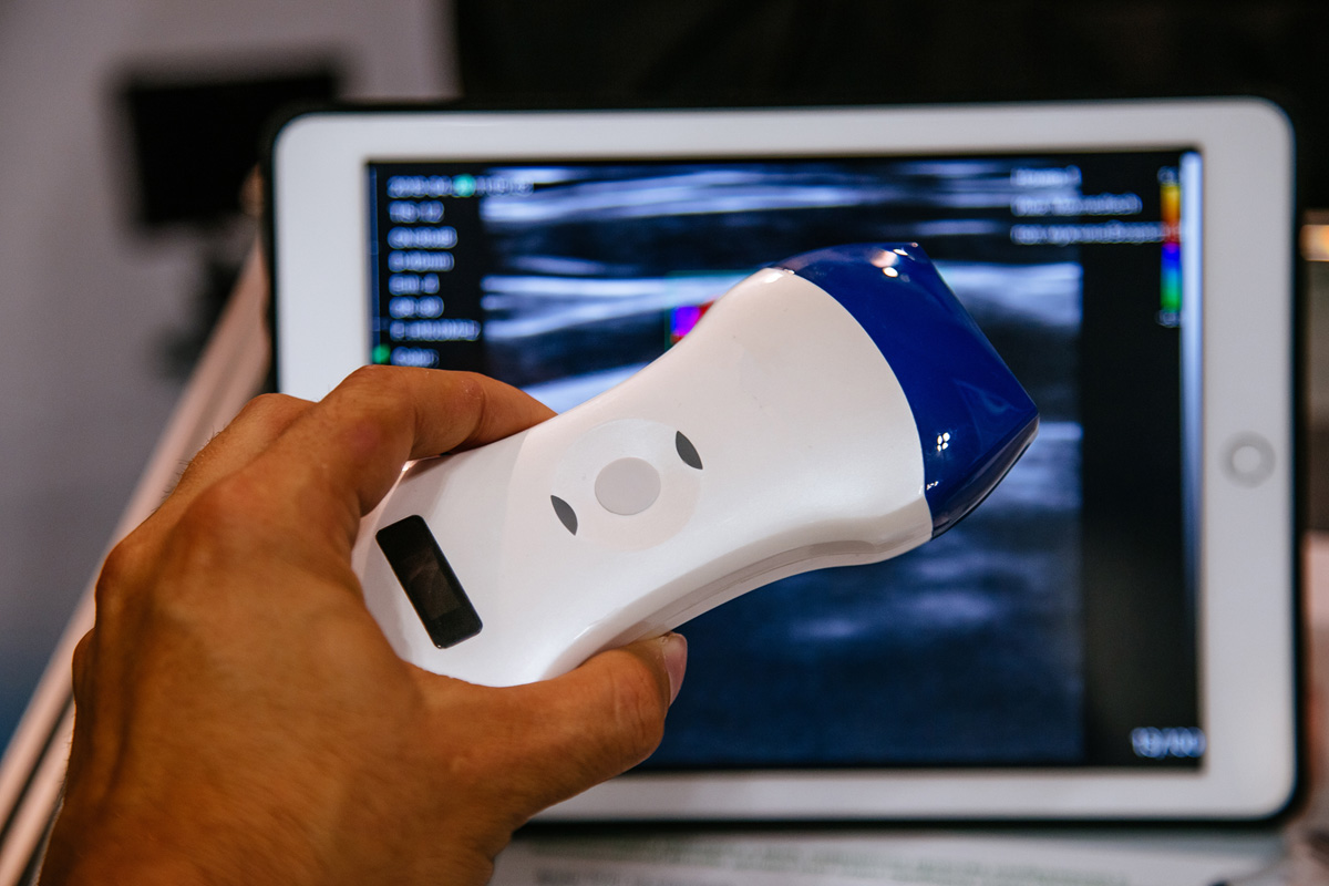
An echocardiogram is the name for an ultrasound of your heart. This exam uses high frequency sound waves to visualize your heart, the inside of your heart, the valve function and the blood flow through your heart.
Patient Preparation
There is no preparation for this exam.

What to Expect During the Exam
Once in the exam room, the technologist will ask you to change into a gown. You will lie on the table on your left side and the technologist will apply three electrodes to your chest to monitor your heart rate during the exam. The technologist will apply warm gel to your chest and use the transducer to create pictures of your heart on the ultrasound machine. The technologist will take several pictures and videos of your heart as measurements are obtained and your blood flow is checked. The technologist will move to different areas of your chest to obtain different information of your heart. You will feel nothing during this exam except for the pressure of the ultrasound probe against your skin. This exam will take 30-45 minutes. Once completed, the exam will be reviewed by our cardiologists and results will be discussed with you during your follow up office visit.
An echocardiogram may be ordered with contrast study. A contrast agent is injected into a vein and is used to highlight certain parts of the heart that may not be visualized well during a regular echocardiogram. Venous access is necessary for this part of the exam and an IV will be placed for the injection of the contrast agent.
An echocardiogram may be ordered with a bubble study. A bubble study is a saline injection that can show if there is an abnormal connection between the right and left side of the heart. Venous access is necessary for this part of the exam and an IV will be placed for the saline injection.

